
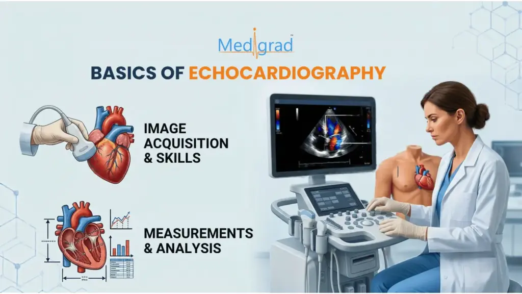
Echocardiography is an important non-invasive imaging modality for assessing cardiac structure, function and blood flow. For doctors, understanding the basics of echocardiography can provide a stronger foundation for evaluating cardiac anatomy, ventricular function, valves and hemodynamics.
Modern echocardiography goes beyond a conventional 2D image. It combines 2D imaging, M-mode, color Doppler, pulsed-wave Doppler, continuous-wave Doppler and advanced techniques to provide structural and functional information about the heart.
For doctors developing their cardiovascular imaging knowledge, the key is not simply knowing what an echocardiogram shows, but understanding how images are acquired, what measurements mean and how findings are interpreted in clinical context.
What Are the Basic Echocardiography Techniques?
A foundation in echocardiography starts with understanding the main imaging modes and how each contributes to cardiac assessment.
1. Two-Dimensional Echocardiography
2D echocardiography provides moving cross-sectional images of the heart. Standard views such as the parasternal long-axis, parasternal short-axis and apical views allow assessment of cardiac chambers, valves and wall motion.
Developing the ability to obtain and recognize standard echo views is one of the fundamental echocardiography skills for doctors.
2. M-Mode Echocardiography
M-mode records motion along a single ultrasound beam over time. Its high temporal resolution can be useful for evaluating movement and making selected cardiac measurements.
3. Doppler Echocardiography
Doppler techniques provide information about blood-flow velocity and direction.
Doctors commonly encounter:
- Color Doppler – visualizes blood flow over the 2D image.
- Pulsed-wave Doppler – measures flow velocity at a selected location.
- Continuous-wave Doppler – measures high velocities along the ultrasound beam and is particularly useful in assessing certain valvular lesions.
Understanding the principles of Doppler is essential for interpreting hemodynamic information obtained during an echocardiographic examination. The accuracy of Doppler measurements also depends on appropriate technique and understanding of flow dynamics.
Essential Echocardiography Skills for Doctors
Learning echocardiography involves more than memorizing normal values. Doctors need to develop a combination of image acquisition, measurement, interpretation and clinical reasoning skills.
Important foundational skills include:
- Recognizing standard echocardiographic views
- Understanding cardiac anatomy in different imaging planes
- Optimizing image quality and gain settings
- Identifying cardiac chambers and valves
- Understanding basic Doppler principles
- Assessing ventricular systolic function
- Recognizing common valvular abnormalities
- Understanding basic hemodynamic measurements
- Identifying abnormal wall motion
- Correlating echocardiographic findings with the clinical picture
- Recognizing when additional imaging or specialist assessment may be required
What Measurements Are Important in Echocardiography?
Echocardiographic measurements help transform visual observations into quantitative information about cardiac structure and function.
Depending on the examination and clinical question, doctors may encounter measurements related to:
- Chamber dimensions and volumes
- Left ventricular systolic function
- Right ventricular function
- Wall thickness
- Doppler velocities
- Pressure gradients
- Stroke volume and cardiac output
- Diastolic function
- Valvular stenosis and regurgitation
- Pulmonary pressure estimates
What Can Echocardiography Help Assess?
Echocardiography provides information about cardiac structure, function and blood flow, helping doctors assess:
- Cardiac structure: Chambers, ventricular walls, valves, pericardium and great vessels.
- Cardiac function: Ventricular function, wall motion, diastolic filling, cardiac output and hemodynamics.
- Valvular disease: Mitral, aortic, tricuspid and pulmonary valve stenosis or regurgitation.
- Cardiomyopathies and structural abnormalities: Including hypertrophic, dilated and restrictive cardiomyopathies, congenital and other structural heart conditions.
Overall, echocardiography is an important tool for non-invasive cardiovascular assessment and cardiac imaging.
From Basic Echo to Advanced Echocardiography
Once the fundamentals are understood, doctors can progress to more advanced techniques and applications.
These may include:
- 3D echocardiography
- Transesophageal echocardiography
- Stress echocardiography
- Tissue Doppler imaging
- Speckle-tracking echocardiography
- Strain imaging
- Advanced valvular assessment
- Prosthetic valve assessment
- Congenital heart disease imaging
- Advanced hemodynamic assessment
For example, tissue Doppler and speckle tracking can provide additional information about myocardial motion and cardiac mechanics beyond conventional imaging.
The appropriate technique depends on the clinical question, available equipment, operator expertise and the patient’s circumstances.
Why Should Doctors Develop Echocardiography Skills?
For physicians working in internal medicine, cardiology, emergency medicine, critical care, radiology or related clinical settings, echocardiography knowledge can strengthen understanding of cardiovascular structure and function.
The value of learning echocardiography lies in developing a structured approach:
Acquire → Optimize → Measure → Interpret → Correlate clinically
A doctor who understands the fundamentals can better recognize what an echocardiographic finding represents, understand its limitations and communicate more effectively with cardiovascular imaging specialists.
However, competency in echocardiography requires structured education, supervised practice and continued experience. Reading about echo techniques alone is not equivalent to developing practical scanning and interpretation skills.
How Can Doctors Build Echocardiography Expertise?
Doctors can progress from basic concepts to more advanced echocardiography through structured online echocardiography course that combines theoretical knowledge with case-based learning and practical exposure.
For doctors who want to develop deeper expertise in 2D echocardiography, Doppler, cardiac structure and function, valvular disease and echocardiographic interpretation, a structured fellowship can provide a more systematic learning pathway than isolated online resources.
Medigrad Fellowship in Echocardiography
Medigrad’s Fellowship in Echocardiography is designed for MBBS doctors, MD/DNB physicians, radiologists and cardiologists seeking structured education in cardiac ultrasound imaging. The program covers ultrasound physics, cardiovascular anatomy, 2D and 3D echocardiography, Doppler techniques, cardiac diseases and echocardiographic interpretation.
The program combines online academic learning with interactive sessions and a hospital observer ship component. Its curriculum progresses from echo basics and Doppler principles to hemodynamics, valvular disease, ventricular function, cardiomyopathies, congenital heart disease, 3D echo, tissue Doppler and speckle tracking.
For doctors outside India, the online learning component can provide a flexible way to study echocardiography alongside existing professional responsibilities. International doctors should confirm eligibility, practical-training availability and applicable arrangements with Medigrad before enrolment.
Learn how structured expert mentorship elevates clinical scanning skills in our guide: Master the Art of Echocardiography with a Fellowship Program.
Frequently Asked Questions
What are the basics of echocardiography?
The basics include understanding cardiac ultrasound principles, standard imaging views, 2D and M-mode imaging, Doppler techniques, cardiac anatomy, basic measurements and the interpretation of common structural and functional findings.
What skills are needed for echocardiography?
Important foundational skills include obtaining standard views, optimizing image quality, recognizing cardiac structures, applying Doppler techniques, making appropriate measurements and interpreting findings in clinical context.
What is the difference between 2D and Doppler echocardiography?
2D echocardiography primarily provides real-time anatomical and structural images, while Doppler techniques provide information about blood-flow velocity and direction. They are commonly used together during cardiac assessment.
Can MBBS doctors learn echocardiography?
Yes. Doctors can undertake structured echocardiography education after MBBS, depending on the eligibility and scope of the specific program. Medigrad lists MBBS doctors among the eligible backgrounds for its Fellowship in Echocardiography.
Is echocardiography useful for doctors outside cardiology?
Echocardiography knowledge can be relevant to physicians working in areas such as internal medicine, emergency care, critical care and other settings where cardiovascular assessment is important. The depth of training required depends on the doctor’s role and intended scope of practice.
What should doctors learn after echocardiography basics?
After mastering the fundamentals, doctors can progress to advanced Doppler assessment, valvular disease, ventricular function, hemodynamics, 3D echocardiography, tissue Doppler, strain imaging and other specialized applications.

